Abdominal Pelvic Ultrasound
Ultrasound imaging is extensively utilised to diagnose conditions of the gastrointestinal and pelvic regions. This allows our specialists to capture detailed, real-time views of the liver, gallbladder, spleen, pancreas, bladder, and surrounding organs.
Ultrasound Services: What to Expect
At our hospital, we are committed to providing you with advanced, compassionate diagnostic care. Ultrasound imaging (also known as sonography) is a safe, painless, and non-invasive way for our medical team to view the inside of your body in real-time, helping us deliver an accurate diagnosis without the use of radiation.
How Does an Ultrasound Work?
Ultrasound technology uses high-frequency sound waves—completely undetectable to the human ear—to create detailed images of your internal organs. As these gentle sound waves glide through the body, they bounce off organs and tissues. Our state-of-the-art equipment captures these echoes, instantly translating them into live images on a monitor for our medical team to evaluate.
Common Uses of Ultrasound Imaging
We frequently use ultrasound to examine the abdominal, pelvic, and gastrointestinal organs. It is a vital tool for diagnosing and evaluating conditions affecting the:
Liver and gallbladder
Spleen and pancreas
Bladder and kidneys
Pelvic and reproductive organs
The Benefits & Risks
The Benefits: Because ultrasound uses sound waves rather than ionizing radiation, it is exceptionally safe. It provides our physicians with real-time, moving images, allowing us to see not just the structure of your organs, but how they are actively functioning.
The Risks: There are no known risks or side effects associated with diagnostic ultrasound. If you are already experiencing tenderness or pain in your abdomen or pelvis, you may feel mild, temporary discomfort as the technician applies gentle pressure to get a clear image.
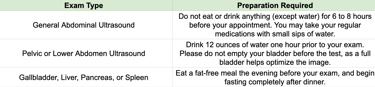
How to Prepare for Your Ultrasound
Your preparation depends on the specific type of ultrasound your physician has ordered. Following these guidelines ensures we get the clearest possible images.
What to Expect During the Procedure
Our team prioritizes your comfort and privacy every step of the way.
Preparation: You may be asked to remove jewelry or clothing that could block the scan. Please note: Your comfort is our priority. If you would prefer a medical chaperone to be present during your exam, simply let our staff or your sonographer know.
Positioning: You will lie down on a comfortable exam table, and we will provide a medical drape sheet to ensure your privacy while exposing only the area being imaged.
The Scan: A certified sonographer will apply a warm, water-soluble gel to your skin. This gel eliminates air pockets and helps transmit the sound waves. They will then gently move a handheld device (a transducer) over the area.
Timing: The entire process is quick and efficient, typically taking just 15 to 20 minutes. Once finished, we will wipe away the gel, and you can return to your day.
Note: If at any point during the scan you experience pain, please tell your technician immediately.
Factors That Can Affect Your Results
To ensure the highest quality images, our team works to minimize any interference. However, certain factors can occasionally make it difficult to get a clear picture, including:
Severe obesity
Food remaining in the stomach
Excess intestinal gas
Your Next Steps: After the Test
Once your exam is complete, one of our board-certified radiologists will review and interpret your images.
Your doctor will discuss the results with you directly. Depending on your preference and the urgency, we will reach out via phone, or during a follow-up appointment. If needed, your physician will discuss next steps, further testing, or tailored treatment plans.